Correlation between P53 and Some Interleukins and Study the Role of P53 Gene in Breast Cancers
Mayada Al-khafaji* and Zaedoon Monaam
Department of Medical Lab Techniques, College of Health and Medical Techniques, Gilgamesh University, Baghdad, Iraq
Published Date: 2025-06-23Mayada Al-khafaji* and Zaedoon Monaam
Department of Medical Lab Techniques, College of Health and Medical Techniques, Gilgamesh University, Baghdad, Iraq
- *Corresponding Author:
- Mayada Al-khafaji Department of Medical Lab Techniques, College of Health and Medical Techniques, Gilgamesh University, Baghdad, Iraq E-mail: mayada.h.mahdi@gu.edu.iq
Received date: December 16, 2024, Manuscript No. IPRDDT-24-20156; Editor assigned date: December 19, 2024, PreQC No. IPRDDT-24-20156 (PQ); Reviewed date: January 03, 2025, QC No. IPRDDT-24-20156; Revised date: June 16, 2025, Manuscript No. IPRDDT-24-20156 (R); Published date: June 23, 2025, DOI: 10.36648/2380-7245.11.2.202
Citation: Al-Khafaji M, Monaam Z (2025) Correlation between P53 and Some Interleukins and Study the Role of P53 Gene in Breast Cancers. J Rare Disord Diagn Ther Vol:11 No:2
Abstract
The p53 gene, which is also referred to as a tumor suppressor and the custodian of the genome, is essential for preserving genomic stability in the nucleus of every cell. This research aimed to study the correlation between interleukins and F53 and investigates the potential link between p53 and the development of cancerous lesions, with a particular focus on breast cancer. Sixty samples were collected, comprising. Forty breast cancer specimens and twenty control samples from healthy individuals. The breast cancer samples were obtained from patients at Medical City Hospital, Baghdad. Healthy control samples were collected from volunteer students at the Gilgamesh University. This study employed quantitative Real-Time Polymerase Chain Reaction (RT-PCR) to specifically assess the expression levels of the p53 gene, and ELISA to determine the levels of (IL-2, IL-8 and P53 titer). The analysis revealed that IL-2 and IL-8 showed increase (10.6 ± 3.2 pg/ml, 25.7 ± 4.5 pg/ml) in breast cancer patients compared with control (2.8 ± 1.9 pg/ml, 8.3 ± 1.0 pg/ml), while p53 showed decrease in patients (129.7 ± 55.9 pg/ml) compared with control. Eleven of the 15 breast cancer samples used for RT-PCR exhibited significantly lower p53 expression. These findings suggest a potential correlation between downregulation of p53 expression and breast cancer development. The results showed a significant difference in p53 expression between controls and patients (P>0.05).
Keywords
F53; RT-PCR; Interleukin; Lesions
Introduction
Breast cancer is the most frequently diagnosed malignant tumor in women and the leading cause of cancer-related death worldwide. Globally, the prevalence of breast cancer is steadily rising [1]. The immune system is shielded from cancer initiation through cancer immunity. The humoral immune system (B lymphocytes) and cellular immune system (T lymphocytes) are two defense mechanisms against cancer cells. Cellular immunity is crucial, even though it works against tumor cells. Th1 cells produce the lymphokine Interleukin-2 (IL-2), which is derived from T lymphocytes (T-helper 1) [2].
The chemokine Interleukin-8 (IL-8) has major potential as a prognostic and/or predictive biomarker of cancer. It plays an autocrine and/or paracrine role in tumor generation. IL-8 may have a special function in breast cancer, primarily driven by the expression of Human Epidermal Growth Factor Receptor 2 (HER2) and Estrogen Receptor (ER) [3]. Both malignant and stromal cells generate interleukin-8, which is released in response to endothelial cells and monocytes. Previous research has indicated that IL-8 promotes cell invasion, metastasis, and angiogenesis, which accelerates the growth of breast cancer cells [4].
The fields of immunology and cancer virus research have been used to create the p53 field. It followed the trajectory of cancer research over the last 40 years. Immunotherapy is now the mainstay of cancer treatment, and numerous indications point to the function of the p53 protein as an antigen in adaptive immune responses as well as in the control of the innate immune system. As a component of the innate immune system, p53 gene and protein are crucial for the surveillance of repetitive DNA and RNAs, senescence, aging, and infectious illnesses. In cancers, a mutant form of the p53 protein triggers both a tumor antigen (B-cell antibody response) and tumorspecific transplantation antigen (CD-8 killer T-cell response) [5].
The main function of the p53 protein is tumor suppression. TP53 is responsible for protein expression. This gene is found on the human chromosome 17. It has been suggested that the p53 prevents genomic and phenotypic changes associated with the onset of cancer. This is because of the intricate connections across several signaling pathways that are essential for fundamental biological functions. Apoptosis, autophagy, cell division, immunological response, and Tumor Microenvironment (TME) modulation are some of these mechanisms [6,7].
Consider the following figure. When specific DNA response factors or elements are bound to the wild-type p53 protein, a wide variety of genes are expressed. This results in protection against the onset and spread of cancer [8,9].
A TP53 gene mutation has been found in almost half of human breast cancer cases. In the event of DNA damage, wild- type p53 inhibits cell reproduction until the damage is fixed. Consequently, the spread of cells with faulty DNA ceases. No cancer phenotypes were observed. TP53 mutations affect the cell cycle. These mutations result in loss of cellular regulation during cell division. This causes faulty DNA to pass to the progeny. Consequently, malignant cells arise. Wild-type TP53 was translated into p53. These transcription factors play critical roles in coordinating several cellular reactions. These reactions include cell cycle arrest, differentiation, DNA repair, senescence, cell death, and metabolism. This results in the activation of the biological mechanisms that suppress cancer progression. The "guardian of the genome" is one such response, as genomic integrity is maintained.
Aim of the study The aim of this study investigate the expression of p53 gene in BC patients, and study the immune parameter levels in BC patient and study the correlation between them (Figure 1).
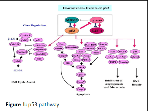
Figure 1: p53 pathway.
Materials and Method
Sample collection
Forty blood samples were obtained from breast cancer patients at the Emergency Center of Medical City Hospitals in Baghdad, and 20 samples were obtained from healthy people between January 2024 and March 2024, patients aged ranged from 35 to 76 years.
Each patient provided five milliliters of blood, which was divided into two milliliters in a gel tube without an anticoagulant, and allowed to clot at room temperature. The blood was then centrifuged at 2000x for ten minutes. Serum samples were kept at -20°C until use for Enzyme-Linked Immunosorbent Assay (ELISA), and 3 ml kept in an Ethylenediaminetetraacetic Acid (EDTA) tube with anticoagulant at -20 for RNA extraction.
Nclusion of primer sequences: The sequences, their respective locations within the gene, and the NCBI accession number (NM_000546.6) have been clearly provided. Below is Table 1. This information has been referenced in the text under the qRT-PCR methodology section:
The primer sequences used in this study, along with their respective locations and the referenced NCBI accession number (NM_000546.6), are listed in Table 1.
| Gene | Primer | Sequence (5' ? 3') | Location within sequence (Nucleotide) | Reference |
| TP53 | Forward | 5'-AGCTTTGAGGTGCGTGTTT-3' | 701-720 | NM_000546.6 |
| TP53 | Reverse | 5'-CTGTTCCGTCCCAGTAGAT-3' | 851-870 | NM_000546.6 |
Table 1: The primer sequences used in this study for the TP53 gene.
Gene reference and primer location: The primers were designed to target specific regions of the TP53 gene, as indicated by their nucleotide positions within the reference sequence (NM_000546.6). These regions were selected based on their relevance to the gene's expression and its role in breast cancer pathogenesis.
Immunological assay
IL-2, IL-8 and p53 were analyzed by using the ELISA test (SUNLONG, China) according to the kit instructions.
RNA extraction by TRIzol™
RNA extraction:
• TRIzol 0.6 ml was used to suspend the blood.
• A 15-minute incubation period was allowed to ensure complete dissociation of the nucleoprotein complex.
• For lysis, 200 μl of TRIzol™ reagent was added, along with chloroform.
• The mixture was incubated at room temperature for 2-3 minutes.
• After incubation, the sample was centrifuged at 10,000 rpm for 10 minutes.
• The mixture separated into an upper colorless aqueous phase and a lower red phenol-chloroform interphase as a result of this centrifugation.
• The RNA-containing aqueous phase was poured into a fresh tube.
• To precipitate the RNA, 200 μl of isopropanol or absolute ethanol was added to the aqueous phase, followed by incubation at room temperature for 2 minutes.
• The mixture was then centrifuged again at 10,000 rpm for 10 minutes, resulting in the precipitation of total RNA on the filter of the spin column tube.
• The supernatant was discarded.
• 0.5 ml of washing buffer 1 was added to the column to resuspension it.
• Centrifuged the tube for 2 minutes at 10,000 rpm.
• The supernatant was discarded.
• 0.5 ml of washing buffer 2 was added to the column to resuspension it.
• The tube was centrifuged for 2 minutes at 10,000 rpm.
• The supernatant was discarded.
• The column was pre-heated with 75 μl of elution solution and centrifuged for 1 minute at 10,000 rpm.
• The total RNA samples were stored in a deep freezer.
Amplification of specific gene cDNA synthesis:
The cDNA was synthesized using the cDNA ready-to-use kit provided by Bioneer, a company based in Korea.
In this process:
• Eighteen microliters of RNA extract were added to a microfuge tube.
• Two microliters of either hexamer primer (for prokaryotic cells) or oligo dt (for eukaryotic cells) were added and thoroughly mixed.
• The resulting mixture was then incubated in a polymerase chain reaction (PCR) machine under specific conditions: 37°C for 10 minutes, followed by 42°C for 1 hour, and finally 95°C for 5-10 minutes in a single cycle.
• The synthesized cDNA was either immediately utilized as a template for qRT-PCR or stored for long-term preservation at -20°C.
quantitative Reverse Transcription-PCR (qRT-PCR)
Real time PCR amplification as follows:
• Two microliter of cDNA was added to PCR tube.
• One microliter of each primers was added.
• The volume was completed to 20 μl with DNase free distilled water.
• The mixture was mixed well and put in the qPCR machine. Table 2 reveals the PCR condition
| Step | Temperature (°C) | Duration | Cycles |
| Initial denaturation | 95 | 3 min | 1 |
| Denaturation | 95 | 15 sec | 40 |
| Annealing | 55 | 45 sec | |
| Extension | 72 | 60 sec |
Table 2: Reveals the PCR condition.
Calculation the fold of gene expression
The fold expression versus the housekeeping gene and control was assessed using the Levak equation by following these procedures.
Ct (Control)–Ct (housekeeping control)=ΔCt (control) Ct (sample)–Ct (housekeeping sample) =ΔCt (sample) ΔCt (sample)
– ΔCt (control) = ΔΔCt
Fold of gene expression = (2¯ΔΔ Ct)
Statistical analysis
The data was analyzed using statistical analysis software, SPSS version 20, for the statistical analysis. The data are shown as mean ± SD. T-test and correlation were used to perform the statistical analysis of the data. The threshold of significance was set at P<0.05.
Result
The total number of a subject that participate were 60 (40 patients and 20 control). Thirteen (32.5%) patients belonged to the 30-44 age group, seventeen (42.5%) to the 45-55 age group, and ten (25%) to the 56-70 age group. According to BMI, the Table 3 explains the number of breast cancer women with BMI, were 5 (12.5%) women underweight (<18.5), 12 (30%) women normal weight (18.5–24.9), 14 (35%) overweight (25–29.9), and 9 (22.5%) with obese (≥ 30).
| Parameters | n | % | |
| Age (years) | 30-45 | 13 | 32.5% |
| 45-55 | 17 | 42.5% | |
| 56-70 | 10 | 25% | |
| BMI (kg/m2) | Underweight (<18.5) | 5 | 12.5% |
| Normal weight (18.5–24.9) | 12 | 30% | |
| Overweight (25–29.9) | 14 | 35% | |
| Obese (= 30) | 9 | 22.5% | |
| Total | 40 (100%) | ||
Table 3: Distribution of the of breast cancer women according to ages and BMI.
The results in Table 4 showed a statistically significant upregulation of both IL-2 (10.6 ± 3.2 pg/ml, 2.8 ± 1.9 pg/ml) and IL-8 (25.7 ± 4.5 p g/mL vs. 8.3 ± 1.0 p g/mL) levels when comparing patients with BC to healthy group (p=0.003, p=0.001), respectively (Figure 2).
|
Parameter |
BC patients |
Controls |
P-value |
|
P53 titer |
129.7 ± 55.9 |
175.6 ± 233.8 |
0.02 |
Table 4: Mean levels of interleukins in breast cancer patients compared with control group.
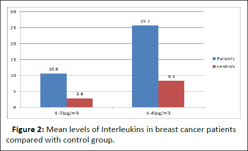
Figure 2: Mean levels of Interleukins in breast cancer patients compared with control group.
Table 5 indicates that women with breast cancer had a lower p53 titer than the control group (Figure 3).
| Interleukins | Patients | Controls | P-value |
| IL-2(pg/ml) | 10.6 ± 3.2 | 2.8 ± 1.9 | 0.003 |
| IL-8(pg/ml) | 25.7 ± 4.5 | 8.3 ± 1.0 | 0.001 |
Table 5: Mean level of P53 titer in breast cancer patients compared with control group.
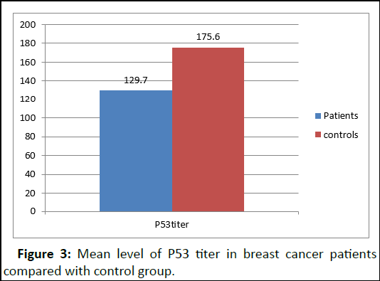
Figure 3: Mean level of P53 titer in breast cancer patients compared with control group.
Figure 4 illustrates the present study's findings, which indicated a positive association (r=0.1) between IL-2 and P53 titer.
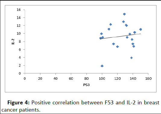
Figure 4: Positive correlation between F53 and IL-2 in breast cancer patients.
The result showed negative correlation between P53 titer and IL-8 (r=-0.2), as showed in Figure 5.
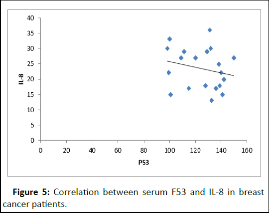
Figure 5: Correlation between serum F53 and IL-8 in breast cancer patients.
The result showed positive correlation between IL-2 and IL-8 (r=0.09), as showed in Figure 6.
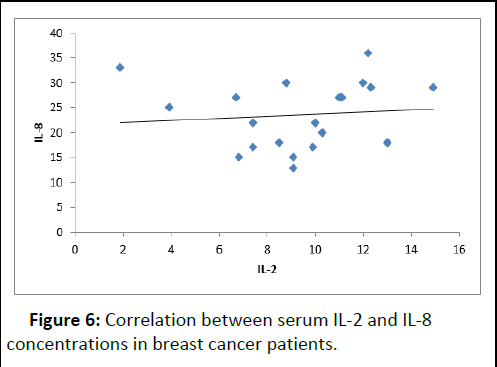
Figure 6: Correlation between serum IL-2 and IL-8 concentrations in breast cancer patients.
Figure 7 based on the study of p53 gene expression involving 15 breast cancer patient samples compared with control samples, the following results were obtained which showed a signi icant difference (P=0.002), as showed in Table 6.
| Sample | Expression fold change2-??Ct |
| Control | 1 |
| Patient 1 | 0.241484 |
| Patient 2 | 0.10083 |
| Patient 3 | 0.291183 |
| Patient 4 | 1.443929 |
| Patient 5 | 0.200267 |
| Patient 6 | 0.334678 |
| Patient 7 | 0.10822 |
| Patient 8 | 0.450678 |
| Patient 9 | 0.200633 |
| Patient 10 | 0.350892 |
| Patient 11 | 1.233658 |
| Patient 12 | 0.1532 |
| Patient 13 | 1.199637 |
| Patient 14 | 0.400539 |
| Patient 15 | 1.608744 |
Table 6: p53 gene expression in Breast cancer patients compared with control.
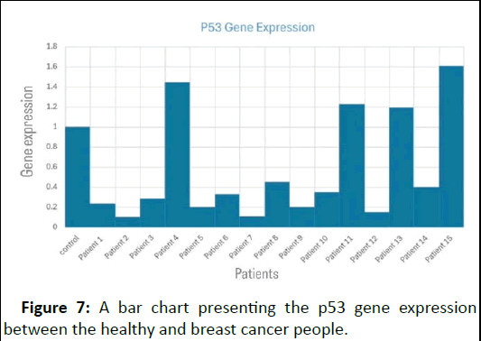
Figure 7: A bar chart presenting the p53 gene expression between the healthy and breast cancer people.
Discussion
The results of the current study are consistent with those of an Iraqi study by Al-Saady, who found that the BMI of breast cancer patients increased significantly. Four main reasons account for the correlation between obesity and breast cancer:
• Metabolism of sex hormones.
• Obese people have lower levels of circulating adiponectin,
• A growth factor that promotes the proliferation of ERα- positive cells.
• Deregulated insulin signaling and chronic low-grade inflammation.
These findings indicated a higher incidence of breast cancer in patients aged 45–54 years, which is consistent with the findings of Khalil et al. and Heer et al. The highest incidence of illness was observed in women aged >48. Since obesity causes the incidence of cancer to increase rapidly with age, it is strongly tied to the age group that had a low incidence when it was younger than 40. The increasing prevalence of breast cancer with age suggests that DNA methylation, a typical aspect of aging, may be one of the causes of increased Breast Cancer (BC) with age. The primary causes of elevated BC in Asia include lifestyle factors, inadequate eating habits, and screening.
Interleukin-2 showed an increase in BC patients compared with the healthy group, which was in agreement with the study of Harjianti et al. and disagreed with Al-Ghurabi et al., which showed a decrease in serum level of IL-2 in BC patients compared with controls. Only Th1 cells can produceIL-2, a lymphokine derived from T-cells. Th1 cells secrete lymphokines that regulate cellular immunity, such as Interferon (IFN-), Tumor Necrosis Factor (TNF-), and IL-2, which activate neutrophils, macrophages, and Cytolytic Cells (CTL). Activation by IL-2, macrophages, neutrophils, and NK cells provides a non-specific defense against tumors. Cell activation can have either cytostatic or cytolytic effects. These cells have the ability to eradicate all varieties of tumor cells because their immunity does not require antibodies or antigen specificity.
A pleiotropic proinflammatory chemokine associated with inflammation, interleukin-8 (CXCL-8), influences angiogenesis and cancer growth, among other cellular processes.
Additionally, this chemokine acts as an autocrine cytokine in tumors. The current study's results showed a substantial increase in IL-8 levels in breast cancer patients compared to the control (P=0.001), a result that agrees with several other studies. According to previous research, IL-8 promotes invasion and metastasis, and is overexpressed in a number of tumor cell types, including those that cause malignancies of the stomach and prostate. Additionally, under some aberrant circumstances, tumor cells release IL-8, which promotes the pro-tumorigenic processes of angiogenesis and cancer cell multiplication.
In line with Kamel et al., the study's mean p53 result showed a significant difference between the two groups, with the control group having a higher mean than the sick group, which is at odds with research conducted by Al-Hassan et al., which demonstrated a substantial rise in the mean serum level of p53- Ab in patients compared to controls. These genes regulate a variety of cellular processes and act as core modules of p53- repressed genes in breast cancer cells by simultaneously responding to genotoxic stress. The expression of p53 in breast cancer ranges from 9% to 69%, The p53 mutation type may be determined by genetic or environmental factors, which may account for the increased expression of p53.
The transfer of combinations of genes producing growth suppressors and immunomodulatory properties may serve as the foundation for novel approaches to enhance tumor regression in vivo. In a transgenic mouse breast model, Pützer et al., examined the efficacy of combination therapy with Advectors expressing p53, a tumor suppressor, and IL-2, an immune stimulator. Murine cells efficiently produced both genes, and Adp53 wt-infected cells underwent apoptosis and showed significantly inhibited cell proliferation. When Adp53 wt and AdIL-2 were combined, the dosage of the latter could be lowered without causing IL-2-related toxicity and without compromising its ability to completely eradicate tumors. The result of Yaun et al., suggests that abnormal p53 expression may be important for modulating angiogenesis, VEGF, and IL-8 expression, which also explains why tumors with high aberrant p53 expression have a poor prognosis.
p53 regulates the expression of genes related to DNA repair, apoptosis (programmed cell death), and cell cycle regulation. This control is essential for preventing damaged DNA-containing cells from proliferating and possibly developing into cancer. Reaction to DNA damage: p53 activity is often elevated in cells in which DNA damage is observed. Next, it encourages DNA repair; if the damage is too great, apoptosis is triggered to prevent the damaged cells from proliferating. Mutation and loss of function: It p53 gene is frequently mutated in various malignancies, including breast cancer. These mutations may result in a lack of tumor suppressor properties, which would allow cells to grow unchecked and develop tumors.
Eleven samples exhibiting low p53 expression were analyzed, and the findings align with numerous studies indicating that diminished p53 levels are frequently correlated with poorer prognosis and altered treatment responses in BC patients. A study featured in BC Research elaborates on how the steadystate level of p53 mRNA is significantly reduced in many breast cancer cases compared to that in normal breast tissue. This decrease in p53 mRNA levels can be attributed to epigenetic modifications such as methylation of the HoxA5 promoter, which consequently impedes p53 expression. International research has emphasized that mutant variants of p53, which may exhibit reduced expression levels, are frequently associated with increased cancer invasiveness and metastasis, further emphasizing the crucial role of p53 in ensuring normal cellular functions.
Four samples exhibiting high levels of p53 expression were obtained from patients, and the results obtained were consistent with numerous studies indicating p53 overexpression. In some breast cancers, p53 protein overexpression often occurs in response to cellular stress or DNA damage. This indicates an underlying mutation in the p53 gene, as mutant forms of the protein tend to be more stable and accumulate within the cells. This accumulation was detectable using Immunohistochemical (IHC) techniques on the tissue samples. Utilizing p53 IHC as a diagnostic tool not only aids in identifying the mutational status of TP53 but also serves as a prognostic marker. High p53 expression levels, as detected by IHC, are frequently associated with high-grade tumors and may correlate with poor outcomes in patients with breast cancer. Tumors exhibiting intense p53 staining are often high-grade and display a negative estrogen receptor status, underscoring the link between p53 overexpression and aggressive tumor characteristics.
Conclusion
The results showed that IL-2 and IL-8 are important biomarkers for prognosis and diagnosis and have a significant influence on the development of breast cancer. They are highly expressed in cancerous cells, and because of their critical functions in the pathogenesis of cancer, they may prove to be attractive targets for cutting-edge treatments aimed at treating breast cancer. TP53 is mutated in approximately half of all human malignancies, including breast cancer. As human cancers frequently have p53 deficiency, this protein is a great choice for cancer treatment.
References
- Smolarz B, Nowak AZ, Romanowicz H (2022) Breast cancer epidemiology, classification, pathogenesis and treatment (review of literature). Cancers 14:2569
[Crossref] [Google Scholar] [PubMed]
- Fasoulakis Z, Kolios G, Papamanolis V, Kontomanolis EN (2018) Interleukins associated with breast cancer. Cureus 10:e3549
[Crossref] [Google Scholar] [PubMed]
- Todorovic-Rakovic N, Milovanovic J (2013) Interleukin-8 in breast cancer progression. J Interferon Cytokine Res 33:563-570.
[Crossref] [Google Scholar] [PubMed]
- Al-Hasso IK (2024) Assessment of serum soluble toll-like receptor-4 and interleukin-8 as biomarkers in patients with breast cancer. Al-Rafidain J Med Sci 6:167-171
- Levine AJ (2020) P53 and the immune response: 40 years of exploration-a plan for the future. Int J Mol Sci 21:541
[Crossref] [Google Scholar] [PubMed]
- Hafner A, Bulyk ML, Jambhekar A, Lahav G (2019) The multiple mechanisms that regulate p53 activity and cell fate. Nat Rev Mol Cell Biol 20:199-210
[Crossref] [Google Scholar] [PubMed]
- Timofeev O (2021) Mutant p53 in cancer progression and personalized therapeutic treatments. Front Oncol 11:740578
[Crossref] [Google Scholar] [PubMed]
- Levine AJ (2019) The many faces of p53: Something for everyone. J Mol Cell Biol 11:524-530
[Crossref] [Google Scholar] [PubMed]
- Harris SL, Levine AJ (2005) The p53 pathway: Positive and negative feedback loops. Oncogene 24:2899-2908
[Crossref] [Google Scholar] [PubMed]

Open Access Journals
- Aquaculture & Veterinary Science
- Chemistry & Chemical Sciences
- Clinical Sciences
- Engineering
- General Science
- Genetics & Molecular Biology
- Health Care & Nursing
- Immunology & Microbiology
- Materials Science
- Mathematics & Physics
- Medical Sciences
- Neurology & Psychiatry
- Oncology & Cancer Science
- Pharmaceutical Sciences